Consolidation in the left apical region Probably due to exacerbated COPD with infective etiology
General medicine :
Here we discuss our individual patient's problems through series of inputs from available global online community of experts with an aim to solve those patient's clinical problems with collective current best evidence based inputs.
This E log book also reflects my patient-centered online learning portfolio and your valuable inputs on the comment box is welcome."
I've been given this case to solve in an attempt to understand the topic of "patient clinical data analysis" to develop my competency in reading and comprehending clinical data including history, clinical findings, investigations and come up with a diagnosis and treatment plan.
Case:
-A 51 year old male patient resident of Miryalguda , farmer by occupation ,presented with a chief complaint of
1. Fever since 10 days
2. Cough with sputum since 10 days
3. Shortness of breath since 7 days
-History of present illness:
Patient was apparently asymptomatic 10 days back then developed following symptoms
Fever which was insidious in onset and it was associated with chills and rigors with diurnal variation which was more during the night and was relieved on medication
He then developed Cough which gradually progressed more during the nights and associated with sputum. It aggrevated during exposure to colder climates .The sputum was scanty and yellow which was non foul smelling (most probably bacterial infestation).
Cough was associated with Chest pain which was non radiating in nature and aggrevated on lying down relieved on sitting upright
He later developed Dyspnea which went on to interfere his daily activities Grade 3 according to MMRC.
-Past history
-No history of Asthma ,Diabetes Mellitus ,Hypertension ,Epilepsy ,siezures
TB : 5 yrs back and was treated with anti tubercular drugs.
-Family history-Not relevant
-Personal history :
Appetite-normal
Sleep: inadequate
Bowel and bladder- regular
Diet: Mixed
No food or drug allergies
Addictions : smoking since 40 yrs ( 3 to 4 cigarettes a day,smoking index-(no.of cigarette*years) -3*40-120.Pack years-6
-Differential Diagnosis
Pneumonia
TB
COPD
-General Examination :
Patient was conscious coherent and cooperative
undernourished,under built
-Vitals
Pulse- 84 bpm, Regular ,Normal volume
Bp -100/70 mm hg
Respiratory rate -24 cpm

-On physical examination
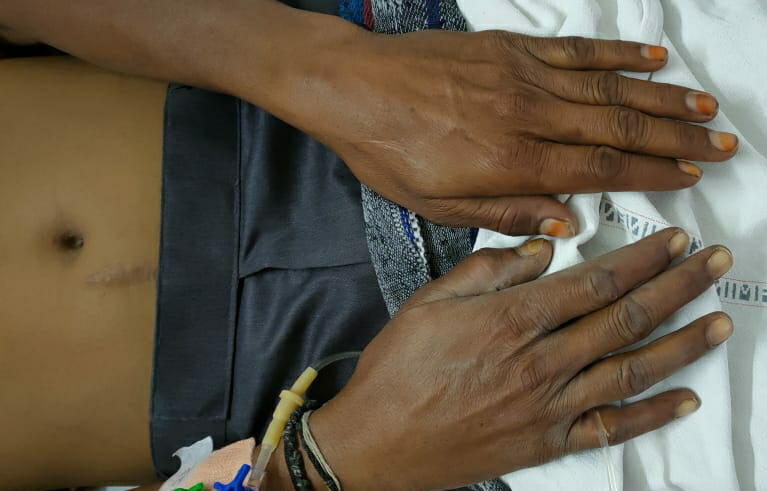
There is no Pallor
Icterus
Cyanosis
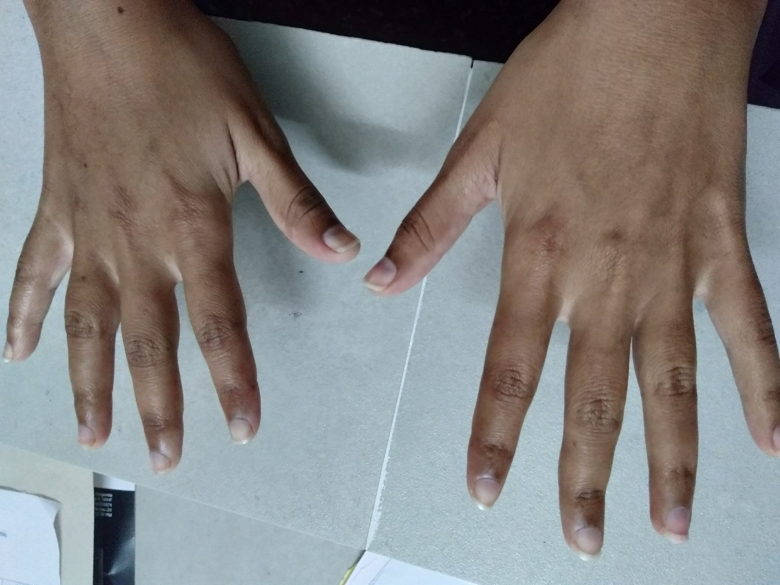
Clubbing
Lymphadenopathy
Edema
Systemic examination
RESPIRATORY
-Upper respiratory tract examination
Nostrils : Normal
Nasal septum: No deviated nasal septum
Nasal polyps: No nasal polyps
Tonsils :No enlarged tonsils
Posterior pharyngeal wall appears to be normal
-Inspection of chest
Shape and symmetry :Elliptical and symmetrical

Spine: central
Trachea :central in position
Respiratory movements - decreased on both sides
Breathing pattern - normal
No visible pulsations
No visible scars or sinuses
PALPATION OF CHEST
- Spine is central
-Trachea is central

-Dimensions AP 16.5 cm
Transverse 23.5 cm
-Chest expansion -decreased

-Vocal fremitus -was increased on left infra clavicular and mammary area

-Apex beat was felt on 5 th intercostal space medial to Mid clavicular line
PERCUSSION OF CHEST

-On purcussion dull note was heard on
-Left infra clavicular
-Left mammary
-Left infra scapular
AUSCULTATION ON CHEST
-bronchial breath sounds are heard
-There was an Increased vocal resonance and crepitations on left infra clavicular and mammary area.
CVS :
Normal S1 S2 heard
No murmurs
Apex beat felt on 5 th intercoastal space
CNS:No focal deficits seen
INVESTIGATIONS


Provisional diagnosis-
Consolidation in the left apical region Probably due to exacerbated COPD with infective etiology
